- Does radiographic pathology feel disconnected from your daily imaging work-like a subject you studied for the exam but never fully applied to the patients, protocols, and clinical decisions you face every shift?
- Have you ever wondered how pathology knowledge, technique modification, modality selection, AI-assisted detection, post-COVID findings, and the latest ARRT content specifications all come together into one complete clinical framework that makes you a better, more confident imaging professional?
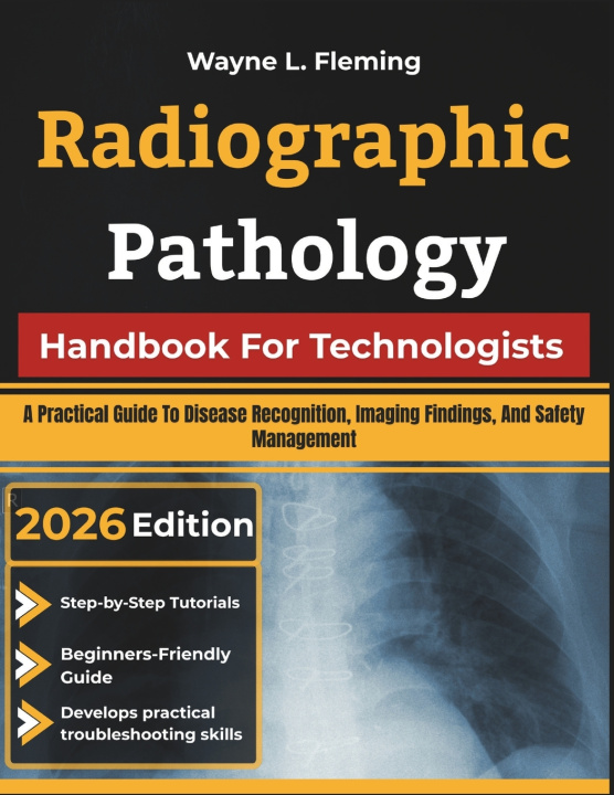
Radiographic Pathology Handbook For Technologists is your complete, body-system-by-body-system guide to understanding, recognizing, and applying pathology knowledge across every major imaging modality-from plain film and CT to MRI, ultrasound, nuclear medicine, and beyond.
Inside This Handbook, You Will Learn How to:
- Master the foundational language of disease-etiology, pathogenesis, cellular injury, necrosis, and neoplasia-and connect each concept directly to the imaging findings it produces
- Apply additive and subtractive disease principles to real-time technique modification for every body system and every pathological condition you encounter
- Recognize the full spectrum of skeletal pathology-fracture classification, metabolic bone disease, arthritis, bone tumors, osteomyelitis-and understand how photon-counting CT is redefining what bone looks like at ultra-high resolution
- Identify lobar, bronchopneumonia, interstitial, and atypical pneumonia patterns across X-ray and CT, and recognize post-COVID pulmonary pathology and AI-assisted chest radiography alerts with confidence
- Assess cardiovascular pathology from cardiomegaly and heart failure to aortic dissection, pulmonary embolism, and post-COVID myocarditis and pericarditis on CT angiography and MRI
- Characterize urinary system pathology from hydronephrosis and pyelonephritis through renal cell carcinoma, urolithiasis by stone type, and bladder malignancy-and implement contrast safety protocols that protect patients with renal disease
- Interpret CNS pathology including ischemic and hemorrhagic stroke, traumatic brain injury, brain tumors, spinal cord compression, multiple sclerosis, and cerebral abscess-and understand how AI stroke triage tools and photon-counting CT are transforming neuroimaging in 2026
- Apply trauma imaging protocols across every body region-polytrauma pan-scan CT, chest and abdominal trauma grading, pelvic ring classification, C-spine clearance, and the imaging recognition of non-accidental trauma in children
- Deploy emerging technologies intelligently-photon-counting CT spectral imaging, deep learning CT reconstruction, accelerated MRI protocols, handheld POCUS, hybrid PET-CT and PET-MRI-with the pathology knowledge that makes each technology clinically meaningful
- Prepare for the 2026 ARRT Radiography examination with confidence-including scenario-based question formats, hotspot image questions, and cross-system pathology integration aligned to the updated content specifications
More Than a Textbook-A Clinical Reference Built for the Real World
This book is not a passive review of conditions and their definitions. It is a working clinical guide for building the integrated, application-ready pathology knowledge that separates excellent radiologic technologists from merely adequate ones.
If you are ready to understand radiographic pathology with clinical depth, real-world precision, and the professional confidence that comes from knowing not just what disease looks like-but why it looks that way-this handbook is your essential guide.
Scroll up and get your copy today-start mastering radiographic pathology the clear, practical, and professionally comprehensive way.